
Center for Collective Development of Scientific Approaches (CCCNP) “Analytical Center for Research and Nanodiagnosis of Materials” was created on the basis of the Institute of Supersolid Materials. V.M. Bakulya NAS of Ukraine.
REGULATIONS ABOUT TsKKNP “ANALYTICAL CENTER FOR ADVANCED NANODIAGNOSTICS OF MATERIALS”
AWARENESS TO THE CENTER
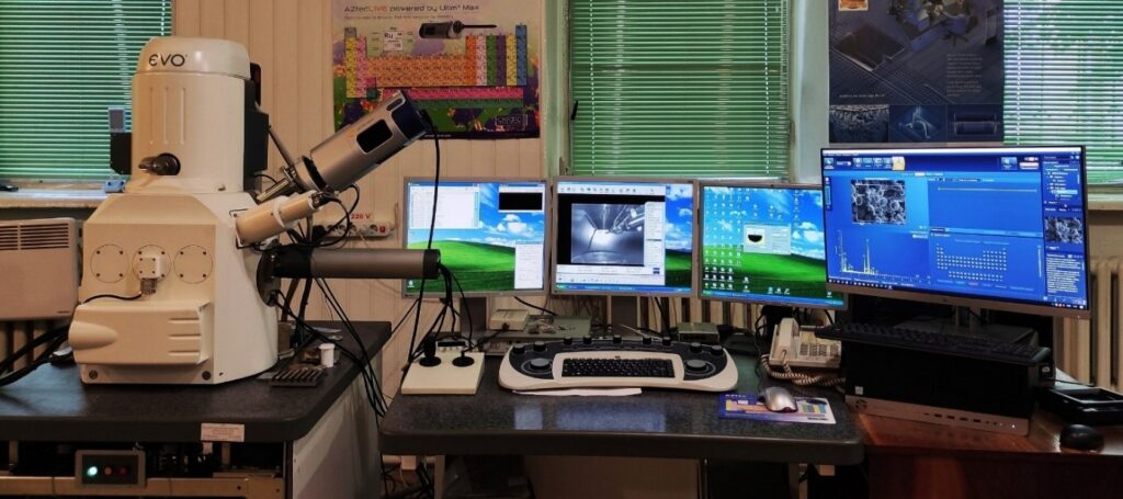
Scanning electron microscope ZEISS EVO 50 XVP (CarlZeiss, Germany)
Main technical characteristics of a scanning electron microscope
Electron microscope ZEISS EVO 50 XVP, equipped with an X-ray spectrum analyzer Ultim Max 100 (Oxford Instruments) and a broken electron diffraction detector. HKL CHANNEL 5. The increase varies between 5x and 1,000,000x.
Basic technical characteristics of a scanning electron microscope:
| Spacious separate buildings at 30 kV: | cathode LaB6 2 nm; |
| cathode W 3 nm; | |
| Fast voltage | range: 0.2 – 30 kV; |
| Strum probe | range: 1 pA – 3 mA; |
| Stability of the electron beam | 0.2%/h. |
Detectors visible on the camera microscope:
– Everhart-Thornley SE secondary electron detector;
– Video camera with IR illumination;
– Highly sensitive 4-quadrant phase CZ BSD detector;
– Detector of secondary electrons for robotic operation in low vacuum and detection of cathodoluminescence topographs.
Analytical devices:
– Energy-dispersive X-ray spectrum analyzer Ultim Max 100 (Oxford), which works with the energy-dispersive spectrometer Aztec live Automate. The separation of the detector is set to 127 eV on the Mn Kal line, 64 eV on the F Kal line, 56 eV on the C Kal line and the transmission speed is 130,000 imp/sec.. Kilkisna the value of the elemental warehouse of the sample from boron to uranium with sensitivity up to 0.1% wt. in local plots 0.1 µm2;
– Detector of spring-impacted electrons HKL Channel 5 (Oxford) for detecting a diffraction pattern from a region greater than 100 nm with specified interplanar distances and traceability of the spring-deformed mill rakes of nanocrystals.
Вимоги до підготовки зразків:
– When a large number of samples are placed in the camera at one time, it is necessary to select samples of the same height;
– In case of high-quality X-ray spectral analysis, the surface roughness should not exceed 1 µm (metalographic surface polish without additional etching of the surface);
– To trace the spring-deformed mill of massive monocrystals and nanocrystals in the solid-state matrix, the surface of the glass is polished, the surface of the damaged ball is removed by a chemical method, electropolishing or otherwise etching.
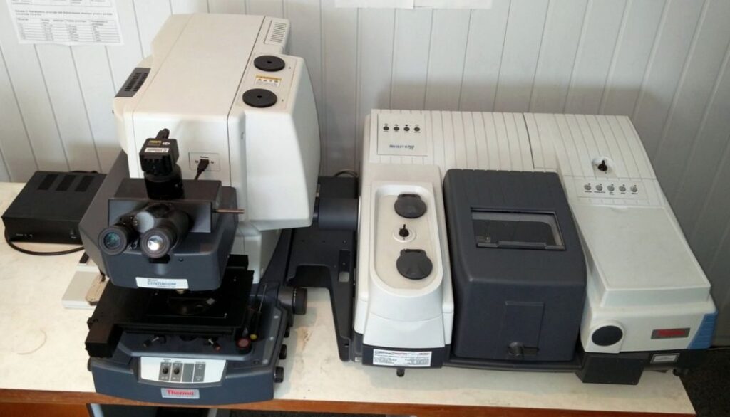
Infrared Fourium spectrometer Nicolet 6700 and microscope Nicolet Continuum (Thermo Fisher Scientific, USA)
Main technical characteristics of the Nicolet 6700 spectrometer:
- Upper spectral range 25000 – 50 cm-1 (0.4 – 200 µm);
- operating ranges that correspond to the same configurations of the detector/light source/detector, near IR region – 25000-8500 cm-1, middle – 7400-600 cm-1, far 600-50 cm-1;
- separate size up to 0.09 cm-1, typical – 1 cm-1;
- diaphragm size for vision – 1 cm;
- system of automatic diagnostics of all units of the device in in-line mode
Quiet modes:
- – transmission/polishing (standard mode) with analytical area 10 mm;
- – beam capacitor (attachment) changes the analytical area to the millimeter;
- – externally damaged internal vibration (ATR prefix) – allows you to implement an express method of investigation, which is especially useful when screwing objects from highly polished materials (from powders to carbide plates;
- – diffuse diffusion (add-on); mirror-image (add-on) with a range of cutouts: 30° – 85°.
Main technical characteristics of the Nicolet Continuum microscope:
- spectral range 7400-500 cm-1;
- Various modes of dimming – transmission/polishing, broken outside internal knocking (ATR), mirror knocking (normal fall);
- The presence of a motorized object table allows for the implementation of mapping patterns over clays or tappings over an analytical area of up to 5×5 µm, typically 100×100 µm.
Ultraviolet-visible spectrophotometer Spekol 1500 (Analytik Jena AG, Nimecchina)
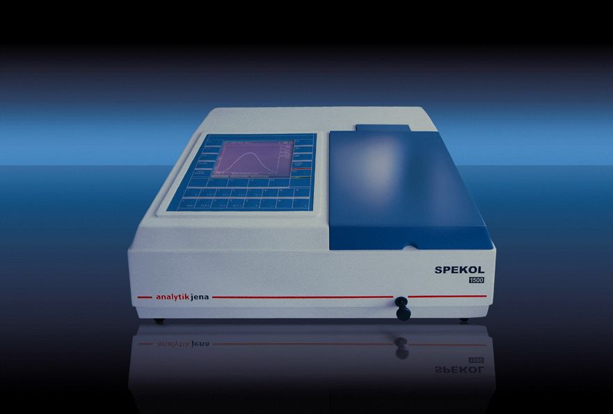
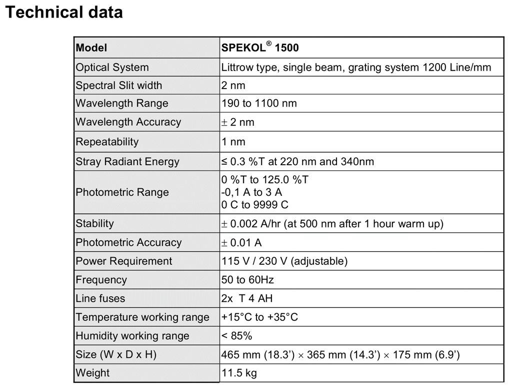
The spectrophotometer is designed to vary the transmittance and optical density of solid, rare and gas-like samples in the ultraviolet and visible spectral range 190-1100 nm. Robotic cleaning can be carried out from a computer, saving the results of calibration and data processing is compiled in the WinASPECT program.